New $180,000 Microscope Used in Cancer, Brain Research
Bruin Notes
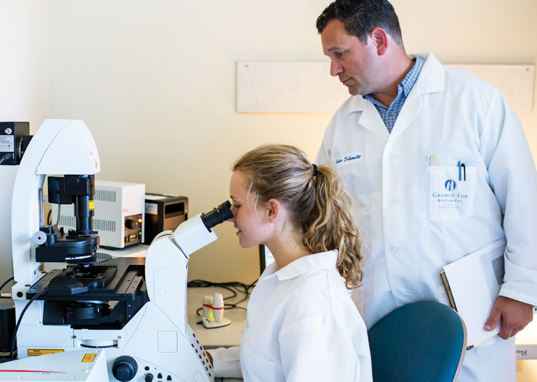
This summer the university’s Department of Biology and Chemistry acquired a state-of-the-art confocal microscope, and already George Fox faculty and students are using it to perform cutting-edge research.
The microscope, manufactured by Leica Microsystems in Germany, typically costs approximately $180,000, but thanks in part to a start-up grant from Leica the purchase was made possible. Confocal microscopy represents a type of microscopy that leverages laser physics to provide high-resolution data to uncover the relationships of molecules within a sample. The new microscope is highly versatile in that it allows users to make comparisons of specific genes, proteins and other molecules in living and developing biological systems in four dimensions, including time.
Already it is being put to good use. Recently, Lael Papenfuse, a biology major and pre-med student, generated a high-resolution image of an aggressive form of breast cancer cells that she and other students study in biology professor John Schmitt’s research laboratory. One of the proteins identified is called “CaM Kinase” and is implicated in cancer growth. “We have never been able to ‘see’ these proteins together in cancer cells before,” says Schmitt. “It’s truly amazing!”
Biology professor Jim Smart and his research students are also using the microscope, in this case to identify key cellular events needed for normal brain development during the periods before and following birth. “Understanding how the brain develops will provide insight into the molecular etiology of brain diseases and disorders like autism, schizophrenia, attention deficit hyperactivity disorder (ADHD) and dyslexia,” says Smart. “The confocal is allowing us to see developmental pathologies in brain tissues that were otherwise undetectable.”